After months of testing with data already published, this January we started the experiments in the laboratory.
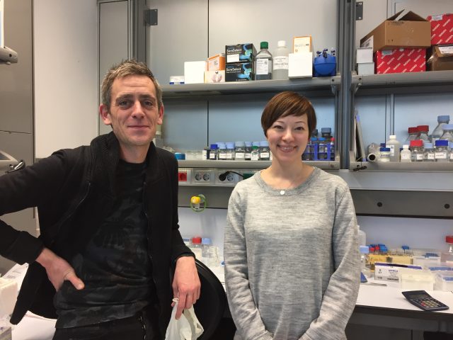
This is an important step: we will extract DNA from the cancer cells that will be analyzed with the game. Yasmina Cuartero and Francois Le Dily are responsible at the CRG for doing the experiments in the lab by applying the Hi-C technique with cells from the T47D line of breast cancer. [You can find more info about the different cell lines we are working with].
We asked them to explain how the technique works and what kind of data it generates.
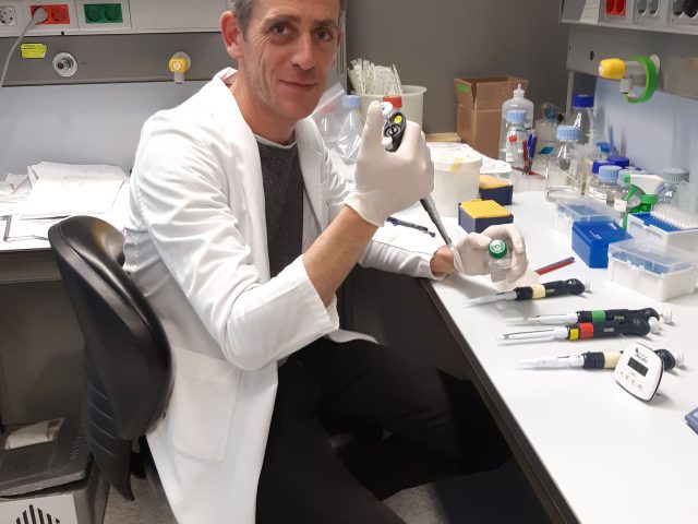
Francois, who works on the response of breast cells to steroids and is an expert in chromatin structure, explains the advantages of this technique: “The Hi-C technique allows to obtain a 3D “photo” of how chromatin (the DNA and associated proteins) is arranged in the cells. This method quantifies the number of interactions between regions of the DNA sequence on a chromosome, which are close to 3D space but may be separated by many nucleotides in the linear genome.
Scientists discovered a few years ago that to understand how a cell works, it is not enough to know what the linear structure of the genes is (i.e. how they are distributed one after the other along the sequence ). This first approach provides only part of the information because DNA is packaged in such a way that there are genes that linearly can be very far away, but in reality they are very close to each other in the 3D space, and from their positions, they interact. It’s about 2 meters of DNA that fold to fit into a nucleus of a few micrometres !
Cancer cells are characterized by genome (the pull of genes) alterations that affect their functioning: this is because their DNA can contain many copies of the same gene (due to an error in the DNA copy), genes that are no longer found in the original position (if compared with structure of a healthy cell), genes that have been “rotated 180 degrees” (therefore its reading fails and do not fulfil its function) or even chromosomes that fuse. In all these cases, there are consequences on proper cellular functioning and can be severe.”
The Hi-C technique makes it possible to find these errors and can be used to reconstruct the genome map of abnormal cells.
On the other hand, Yasmina, an expert in techniques for studying genome structure, gave us some details of how it is applied in the lab. “With the Hi-C technique, we quantify the possible interactions between all pairs of DNA fragments simultaneously. This is done with many cells at once, because, in each cell depending on its life cycle, DNA may be in a different position. The first step is to fix the chromatin with a reagent. Then, with enzymes, 400 pairs of bases are cut in specific parts (based on the knowledge we have for the reference genome of healthy cells) and finally, it binds with another enzyme the pieces that remain close at the 3D level. Once this process is completed, this DNA is extracted to obtaining a series of chimeric fragments (which do not exist in reality in linear form of the genome but are the result of the 3D photo that we have made in the lab ), and that will be sequenced.”
Once it leaves the lab, the sequencing results are passed to a computer program. It generates an array of contacts, containing the information that will be given to volunteers, in the form of a game, to analyze.
This process usually takes several days, but thanks to a laboratory kit offered by ARIMA Genomics, Genigma’s collaborator, we have greatly reduced the times and been able to analyze more than one cell line at a time!
This is how we currently generate the basic data for our game. It is now time to introduce them in our beta version, to test how it works, before launching the app.